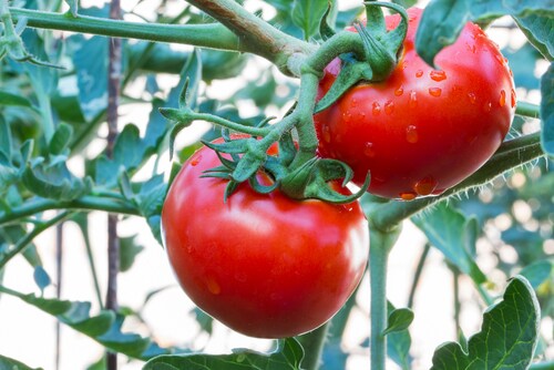
 Single-cell-type proteomics is helpful for studying plant roots, which can contain several populations of unique cells. These cell populations provide information regarding root development and plasticity under various conditions. Researchers Zhu et al. report on a new application of single-cell proteomics using laser capture microdissection (LCM) in tomato seedlings grown in hydroponic tanks.1
The team treated half of the samples with 14.5 μM Al3+ solution to simulate the effects of aluminum toxicity in soil. They harvested the seedlings 10 days after seed germination, at the point when the two cotyledons enlarged and lateral roots began to grow on some of the Al-treated plants. They then fixed the roots and embedded them in optimal cutting temperature (OCT) compound. Using a cryostat, the team collected thin sections, approximately 10-μm thick, with 10 sections per root tip. They mounted the sections onto polyethylene naphthalate (PEN) membrane glass slides (Thermo Scientific) at −20°C.
Using the LCM microscope, the team harvested epidermal and cortical cells (5,000–7,000 cells per tissue type). The LCM cut the single-layer tissues, lifted them from the slides and collected the sample into a capture tube. They then isolated proteins and separated them using sodium dodecyl sulfate polyacrylamide gel electrophoresis (SDS-PAGE), followed by an in-gel tryptic digestion and nano liquid chromatography–tandem mass spectrometry (LC-MS/MS) analysis.
Zhu and colleagues used an Orbitrap Elite mass spectrometer (Thermo Scientific) to serially analyze 10 or 11 individually digested gel fractions.They also used an Orbitrap Fusion Tribrid mass spectrometer (Thermo Scientific) to analyze epidermal samples pooled from five separately digested gel fractions, to avoid the need for pre-fractionation while also allowing for a more rapid scanning rate. Both of the mass spectrometers were coupled with an UltiMate 3000 RSLCnano (Thermo Scientific), using the same LC protocol. After generating MS spectra, the team used Proteome Discoverer software, revision 1.4, (Thermo Scientific) to process raw data and then matched the spectra against the ITAG2.4 tomato genome database to identify proteins contained in each single-cell-type sample.
The researchers identified 1,313 proteins in the Al-treated epidermal cell population and 744 proteins in the Al-treated cortical cell population. They identified 365 proteins in the non-Al-treated epidermal cell population and 745 proteins in the non-Al-treated cortical cell population. Between the Al-treated epidermal and cortical proteomes, 543 proteins overlapped in these two layers of tissues. Between the non-treated epidermal and cortical proteomes, 189 proteins overlapped in these two types of tissues. In both cases, they found a significant portion of the proteome contained proteins unique to the different layers and cell type. Another unique finding was the identification of several important proteins related to Al-induced morphological characteristics of roots. These proteins include the ROOT GROWTH DEFECTIVE 3; the ROOT HAIRLESS 1, required for root hair initiation in Arabidopsis; and the DEFORMED ROOTS AND LEAVES 1 for short root phenotypes, in the epidermal proteins.
These results demonstrate the cell-type-specific proteome is relevant for tissue-specific functions in tomato roots. The authors also note that this technique is still in its infancy with plenty of room for improvement.
Reference
1. Zhu, Y., et al. (2016) “Development of a laser capture microscope-based single-cell-type proteomics tool for studying proteomes of individual cell layers of plant roots,” Horticulture Research, 3, doi:10.1038/hortres.2016.26.
Leave a Reply