More than a century ago, renowned mathematician Johan Radon provided the mathematical framework for tomographic reconstruction. Many years later, in 1968, David DeRosier and Aaron Klug published a milestone Nature paper reconstructing for the first time a three-dimensional bacteriophage tail from its two-dimensional projections.
This marked the dawn of modern electron tomography—a technique that reveals the internal structure of an object through various projections recorded using an electron beam that illuminates the specimen.
Cryo-electron tomography
Today, after decades of developing this method, which included advancements in cryo techniques to obtain intact biomolecules in the hydrated state and achieve conditions for non-destructive irradiation in the electron microscope, cryo-electron tomography achieves unsurpassed levels of resolution.
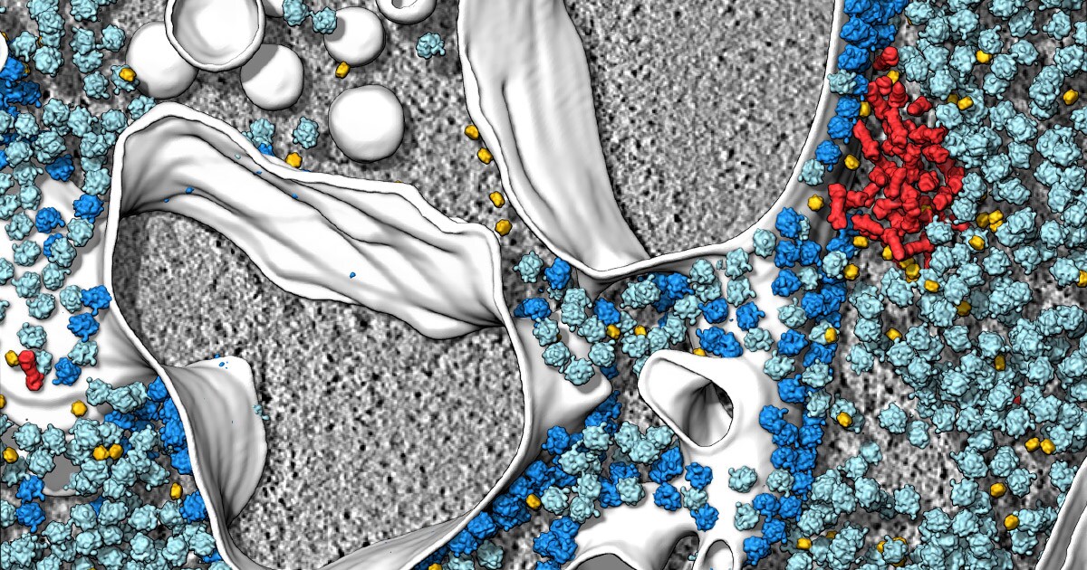
Cryo-electron tomography reveals a phase-separated protein degradation microcompartment where proteasomes (red) cluster at the endoplasmic reticulum membrane (cytosolic ribosomes: light blue; membrane-bound ribosomes: dark blue; Cdc48: yellow; membranes: white; Protein structures obtained by subtomogram averaging and subsequently mapped back into the cellular tomogram). Data courtesy of Dr. Benjamin Engel, Helmholtz Zentrum München.
Bridging the gap between structural biology methods and existing light microscopy techniques, cryo-electron tomography provides three-dimensional imagery and snapshots from the native cell interior.
It enables the study of how proteins and other molecules work together to carry out major processes in a cell, transforming our view of how cells function and giving us new biological insights that no other method provides.
Thanks to technological progress, increased digitization, and transformative advances in the areas of detection, sample preparation, and automation, cryo-electron tomography advanced to what it is today: a soon-to-be fundamental piece in our imaging toolbox with enormous potential in cell biology research.
Explore a curated collection of scientific publications highlighting the use of cryo-electron tomography with our new Cryo-Electron Tomography e-Book.
What’s possible: a new age for cellular biology
I am continuously fascinated by the research results flowing in from across different fields of application as a direct result of cryo-electron tomography.
We’ve learned of cellular structures that were previously difficult to visualize, like degradation microcompartments at the endoplasmic reticulum membrane (2020); large macromolecular assemblies with an intricate architecture, such as nuclear pore complexes, which can now be elucidated in their cellular context (2020); and disease-related ultrastructural changes that are unveiled—most recently, a kinase linked to Parkinson’s disease that was solved by in-situ tomography (2020) or neurotoxic protein aggregates stalling the cellular protein degradation machinery (2018).
Our role at Thermo Fisher Scientific is to help biologists maintain this momentum through continued technological innovation, and to make sure they maximize the far-reaching impacts cryo-EM could have across their research.
Our transmission electron microscopes (TEMs), including the Thermo Scientific Krios G4 Cryo-TEM and Glacios Cryo-TEM, enable higher throughput at increased levels of automation than ever before. At the same time, Cryo-FIB technology, including the Thermo Scientific Aquilos 2 Cryo-FIB, evolved into a game-changing method for sample preparation for tomography—enabling biologists to literally open windows (cryo-lamellae) into functional cellular environments.
We’re excited to also announce our latest innovation in cryo-EM—the Thermo Scientific iFLM Correlative System, a must-have component for the Aquilos 2 Cryo-FIB and our first integrated fluorescence light microscope.
Empowered by cryo-FIB milling, cryo-electron tomography is well on its way to becoming a primary technique for exploring the native cellular environment.
//
Alexander Rigort is a product marketing manager at Thermo Fisher Scientific.
Subscribe Now to receive new Accelerating Microscopy posts straight to your inbox.

How artificial intelligence (AI) improves image analysis workflows in life sciences
Image acquisition is not the end of the story—it is the st...
Read More
Advances in high-resolution cryo-EM at 100 kV
100 kV cryo-TEM enables high-resolution single particle anal... Alex Ilitchev, PhD
Read More
3D Tissue Histology with Light-Sheet Microscopy Enables Nondestructive Analysis of Microglia
3D tissue analysis offers critical benefits for neuroscience... Alex Ilitchev, PhD
Read More
Fragment based drug discovery meets challenging drug targets with high-throughput cryo-EM
Benefits of FBDD in the search for novel therapeutics Frag... Dominic Meusch
Read More
Leave a Reply